
Sup209
Revision of DICOM Conformance Statement
This Supplement provides updates to part PS3.2 of the
DICOM standard, redefining the content and structure of
the DICOM Conformance Statement to better meet the needs
of all user groups, for example service, R&D, testing,
sales, healthcare provider IT personnel.
Comparability is better facilitated for different products' DICOM
functionality by providing essential information in tables.
Ambiguities and inconsistencies will be less frequent between
different vendor documentations.
Web services and security are additionally addressed in the
conformance statement.
A detailed template is provided. Vendors are encouraged to populate
this template for their products. Template-based comparison of
products is advantageous in many situations.
Supplement 209 was voted Final Text and will be incorporated in the
next publication of the standard.
View slideset »

Sup213
2G-RT: Enhanced RT Image
The Supplement addresses imaging within Radiotherapy treatment
sessions and acquiring patient positioning information.
The supplement adds three IODs. Two for supporting projection images
and one IOD supporting acquisition instructions for images and other
artifacts to be used for patient positioning.
The Enhanced RT Image covers the images with a smaller number of
frames, where the per-frame functional group macros are populated for
all frames.
The Enhanced Continuous RT Image covers images which are continuously
acquired, resulting in high number of frames due to a high frame
rate. With frame level attributes not being repeated for each frame
this image type is more efficiently and sparsely populated.
Both IODs represent projection images of the patient geometry in
relation to the treatment device equipment. They may be used to guide
the positioning of the patient in respect to the treatment delivery
device to ensure delivery of the therapeutic dose to the intended
region. They may also be used to verify the position of the patient
when acquired prior, during or after the delivery of the therapeutic
radiation.
The Supplement additionally specifies a new IOD to convey parameters
instructing devices on how to acquire images or other artifacts used
for patient position verification in Radiotherapy treatment delivery
sessions.
RT Patient Position Acquisition Instruction contains the definition of
the procedures, devices, and related parameters to be used for the
assessment and/or verification of the patient position. The technical
parameters can be defined on any level of detail as needed by a
specific device.
Procedures can be paired to represent related operations like e.g. a
paired orthogonal MV and kV image acquisition.
The scope of therapeutic radiation whose position is verified is
specified by referencing SOP Instances identifying objects like RT
Radiation Set IOD of RT Radiation IODs.
Supplement 213 was voted Final Text and will be incorporated in the
next publication of the standard.
View slideset »

Sup230
TLS Security Update 2021
This Supplement adds two new Secure Transport
Connection Profiles and retires several others.
The IETF recently updated the Best Current Practice
document called BCP-195. The new document no longer
allows downgrading to TLS 1.0 or 1.1, which
necessitates DICOM retiring Secure Transport
Connection Profiles that are based on those
protocols.
The new version of BCP-195 is more in
line with DICOM's B.10 Non-Downgrading BCP 195
Secure Transport Connection Profile.
In addition, the Japanese government has modified
their guidelines for "high-security type" devices,
hence the old Extended BCP 195 profile (B.11) is
also now out of date, needs to be retired, and a new
profile created that reflects the new revisions.
Supplement 230 was voted Final Text and will be
incorporated in the next publication of the standard.
View slideset »

Sup229
Photoacoustic Imaging
This Supplement to the DICOM Standard introduces a new IOD and a
new storage SOP Class for encoding and storing photoacoustic
images.
Photoacoustic imaging (PAI) is an imaging modality that enables
imaging optical absorption in biological tissues with acoustic
resolution.
Contrast is generated through absorption by chromophores that
range from intrinsic absorbers such as hemoglobin and melanin to
extrinsic agents such as indocyanine green (ICG) or diverse types
of nano-particles.
Excitation at multiple wavelengths allows the modality to
discriminate individual chromophores. Prospective applications in
the space of clinical imaging range from classification of breast
cancer lesions through screening of sentinel lymph nodes to
assessment of inflammation.
Photoacoustic Imaging is in widespread use in preclinical research
labs and is recently being translated to clinical applications in
first commercial implementations.
Many (but not all) PAI implementations integrate active pulse/echo
ultrasound in a hybrid imaging system to capitalize on
well-established contrast for anatomical information.
The scope of this IOD is the Photoacoustic (PA) images and
processed images that may be derived from a combination of these
PA images.
Complementary images such as pulse/echo ultrasound are represented
by their native DICOM IODs.
Albeit fusing PA images with US images is the presently most
common scenario, the particulars of the fusion are beyond the
scope of this IOD but an example is provided.
This supplement is voted ready to be sent out for Public Comments.
View slideset »

Sup237
Hi-Resolution ECG Waveform
This supplement adds a SOP Class to store high
resolution ECG data for non-cardiology examinations.
In clinical neurophysiology it is common practice
to acquire ECG data together with the routine scalp EEG or
in case of a sleep study.
The added SOP Class is based on the existing
General ECG SOP Class but with fewer constraints. The
General ECG SOP class can store waveform with 16 bits per
sample. The new SOP class permits 32 bits per sample as an
additional high-resolution level.
This supplement is voted ready to be sent out for Public Comments.
View slideset »

Sup228
DICOMweb API for Server-Side Volumetric Rendering
This supplement introduces web services that enable
a user agent to request server-side volumetric
rendering of 3D volumes (Volume Rendering (VR),
Maximum Intensity Projection (MIP) and Multiplanar
Reconstruction (MPR).
The volume is rendered as a 2D representation,
display parameters are applied to achieve the
requested presentation, and lastly, the 2D
representation is encoded into one or more images of
the requested media type and returned in a response
payload to the user agent.
DICOM Web API for Server-Side Volumetric Rendering
is not intended as an alternative to Volumetric
Presentation states, but a complement in enabling
user agents to request a 3D or 3D temporal rendering
without having specify the numerous and complex
parameters to do so.
This supplement considers a basic and an advanced
(3D aware) client scenario.
The basic client, capable of fundamental operations
to select the rendering type, select a rendering
protocol, or to manipulate volumetric view and
transformations.
For the basic client, this supplement focuses on the
20% of requirements that satisfy 80% of the
interoperability needs. It supports pan, zoom,
rotate, set render type, add annotations.
The 3D Aware client, capable of defining and
manipulating the full breadth of parameters
contained within the Volumetric Presentation State
IOD. In this case, capabilities are limited to
Volumetric Presentation State definition and origin
server capabilities. It support more complex
features like color, shading, lightning,
segmentation, cropping, blending and transparency.
Returned rendered object formats include jpg, gif,
png and animated-gif.
This supplement will be further presented to the
base standard before going out for public comments.
View slideset »

Sup234
DicomWeb Storage Commitment
This supplement adds storage commitment
functionality to DICOMweb. This is an extension to
the existing DICOMweb services, mimicking the
storage commitment service that is already available
using DIMSE.
The storage commitment service is typically used
when an image acquisition system wants to free up
storage space for new studies and asks an archive
system of taking over the storage responsibility for
the images previously being sent from the
acquisition device to the archive.
This supplement will be further presented and
discussed in the base standard group before going
out for Public Comments.
View slideset »
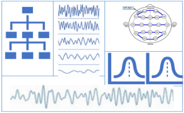
Sup236
Structured Display for Waveforms
This supplement introduces Service Classes for
storage and exchange of presentation information for
DICOM waveform objects by adding Presentation State
and Structured Display IODs and related Context
Groups.
The following IEs and Classes are added to the
standard:
- Waveform Presentation IE
- Montage IE
- SOP Class to store predefined Waveform Montages
- SOP Class to store Waveform Presentation States
- SOP Class to store Waveform Display Settings
- SOP Class to store observations and measurements as annotations
The main use cases that would make use of the new Service Classes are:
- Create a display matching the settings used by the operator during recording
- Create a display matching the settings used by the doctor during review
- Store the annotations and the settings used by algorithms in waveform analysis software
- Time locked annotations with identification of authorship and time of annotation (during acquisition versus post hoc)
In clinical neurophysiology it is important to be able to recreate the presentation of the recorded data as it was displayed during the recording or during review and reporting. This is important for example when activity is noted by the operator during recording and that view needs to be recreated post hoc for specialist review.
This supplement will be further presented and discussed in the base standard group before going out for Public Comments. View details »
View slideset »

Sup238
Assertion Collection
The Assertion Collection IOD persists assertions for
referenced instances and other meta data such as at which
clinical step the assertion was made.
- High-level identification information for the collection
for easy identification.
- Item State definitions (E.g., added, removed, reviewed,
approved) for the referenced Instances on Study, Series,
Instance, or sub-Instance level
- Item State definitions for the Assertion Collection
Instance itself.
The design of the Assertion Collection is agnostic to any clinical domain, and any requires domain-specific information that is modeled by codes, by including specific CIDs or TIDs.
Potential use cases of the Assertion Collection IOD include collection of assertions for instance references during post-acquisition/pre-planning, treatment planning, treatment delivery, pre- or post-treatment quality evaluation.
An Assertion Collection Instance may be used as input for sub-sequent workflow steps, whereas the Assertion Collection IOD only represents a current state and does not include any forward-looking statements about further usage. It is not intended to control any workflow steps, just to represent the outcome.
This supplement will be further presented and discussed in the base standard group before going out for Public Comments. View details »
View slideset »